
Diagnostics
Understanding Dental X-rays and Their Role in Diagnosis
X-rays show what the eye cannot. Here is what they reveal, the main types, and how dentists decide when they are needed.
Dental Planet · · 3 min read
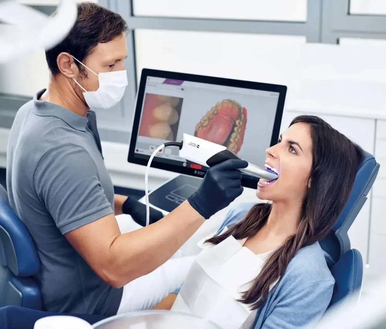
During a dental examination, your dentist can see the surfaces of your teeth and gums. But much of what matters — between the teeth, inside the roots and in the surrounding bone — is hidden from view. Dental X-rays help fill in that picture.
Why dental X-rays are taken
X-rays help dentists identify problems that may not yet cause symptoms, and investigate problems that do. They can reveal:
- Decay between teeth or beneath existing fillings
- Infection at the tip of a tooth root
- Bone levels affected by gum disease
- The position of developing, unerupted or impacted teeth
- Cracks or problems with existing root canal fillings
- Bone volume when planning dental implants
Common types of dental X-ray
Bitewing X-rays
These show the crowns of the upper and lower back teeth together. They are often used to check for decay between teeth.
Periapical X-rays
These show one or a few teeth in full, from crown to root tip, along with surrounding bone. They are often used when investigating a painful tooth or before root canal treatment.
Panoramic X-rays (OPG)
A single wide image of both jaws, all the teeth and nearby structures. Useful for assessing wisdom teeth, overall development and some treatment planning.
3D imaging
In some situations, three-dimensional imaging may be recommended for more detailed assessment. Your dentist will explain if and why this is needed.
What happens during an X-ray
For small X-rays, a sensor is placed in your mouth and held steady while a brief exposure is taken. For panoramic images, you stand or sit while the machine rotates around your head. The process takes only a short time once positioned. Remove items such as earrings, glasses or removable dentures if asked.
How often are X-rays needed?
There is no single schedule for everyone. Dentists consider your history of decay, gum health, symptoms and treatment plans. Someone with a high risk of decay may need X-rays more often than someone with very healthy teeth. X-rays should be taken only when the information is expected to benefit your care.
Safety
Dental X-rays use low doses of radiation, and dentists aim to keep exposure as low as reasonably achievable. Always tell the team if you are, or might be, pregnant, and feel free to ask why a particular X-ray is being recommended.
How X-rays fit with digital scans
Digital intraoral scans capture the surfaces of teeth and gums using light, while X-rays show internal structures. Read how digital dental scanning helps during treatment planning to learn how the two complement each other.
Reading your X-ray with your dentist
X-rays can be hard to interpret without training. Ask your dentist to point out what they are seeing — healthy structures as well as any concerns. Understanding your images can make treatment decisions clearer.
Important to remember
An X-ray is one part of a diagnosis. Your dentist combines it with your symptoms, examination and history before making recommendations. Learn more about our digital scanning and X-ray services, or contact us with any questions.
This information is general and educational. It does not replace an examination or individual advice from a qualified dental professional. Treatment suitability, risks, recovery and results vary from person to person.
Related treatments